About
Dual-Modality Imaging
|
Optical coherence tomography (OCT) is a non-invasive interferometric imaging technique capable of imaging up to 2 mm deep tissue, using backscattered near-infrared light from index of refraction mismatches to create cross-sectional images. Recently, it has been used to image the human colon and rectum with micron-scale resolution. Our in vivo work on the mouse colon with laser-induced fluorescence (LIF) spectroscopy, similar to that which has demonstrated promising capabilities in the human lung and gastrointestinal tract, has shown high sensitivity and specificity in spectrophotometric analysis of distinguishing normal tissue from adenoma. Multiple researchers have shown the ability of LIF techniques to identify cancer and pre-cancer tissue in the human colon using only endogenous fluorophores [Richards-Kortum 1991]. Combining OCT and LIF in one endoscope, with LIF providing information about the biochemical composition and OCT information about plaque boundaries, structure, and thickness, shows a heightened sensitivity to early changes in tumor progression when compared to either modality alone. Previously, we have built ultrahigh resolution (2-5 μm) OCT endoscopes with unfocused LIF and have demonstrated imaging of mouse colon serially over time [Hariri 2006]. A diagram of the setup is shown below. While the optical paths of each modality are quite different proximally, they are combined distally in the sample arm. While OCT requires a focused NIR beam for deep penetration (we use broadband SLD sources with center wavelengths of either 890 or 1300 nm) , LIF resolution can be increased by focusing a UV beam on the same spot. Doing so allows for deep (~2 mm) and shallow (~200 μm at 325 nm excitation) penetration with the two modalities, respectively, as well as for heightened specificity and sensitivity for precancerous adenoma detection.
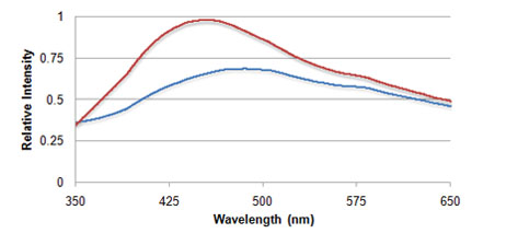
In order to further increase our ability to distinguish normal tissue from adenoma, we have built a dual-modality endoscopic instrument that produces high-resolution OCT images and fluorescence spectra during in vivo imaging of the mouse colon. Images below, taken from a diseased mouse show a 30 mm OCT scan and LIF spectra taken across healthy tissue and adenoma.
References Richards-Kortum, R., Rava, R. P., Petras, R. E., Fitzmaurice, M., Sivak, M. and Feld, M. S., “Spectroscopic diagnosis of colonic dysplasia,” Photochem. Photobiol. 53(6), 777-786 (1991). Izatt, J. A., Kulkarni, M. D., Yazdanfar, S., Barton, J. K., and Welch, A. J., “In vivo biodirectional color Doppler flow imaging of picoliter blood volumes using optical coherence tomography,” Opt. Lett. 22, 1439-1441 (1997). Hariri, L. P., Tumlinson, A. R., Besselsen, D. G., Utzinger, U., Gerner, E. W. and Barton, J. K., “Endoscopic optical coherence tomography and laser-induced fluorescence spectroscopy in a murine colon cancer model,” Lasers Surg. Med. 38, 305-313 (2006). Barton, J. K., Guzman, F. and Tumlinson, A. R., “Dual modality instrument for simultaneous optical coherence tomography imaging and fluorescence spectroscopy,” J. Biomed. Opt. 9(3), 618-623 (2004). Tumlinson, A. R., Hariri, L. P., Utzinger, U. and Barton, J. K., “Miniature endoscope for simultaneous optical coherence tomography and laser-induced fluorescence measurement,” Appl. Opt. 43(1), 113-121 (2004). |