About
Falloposcope Overview
|
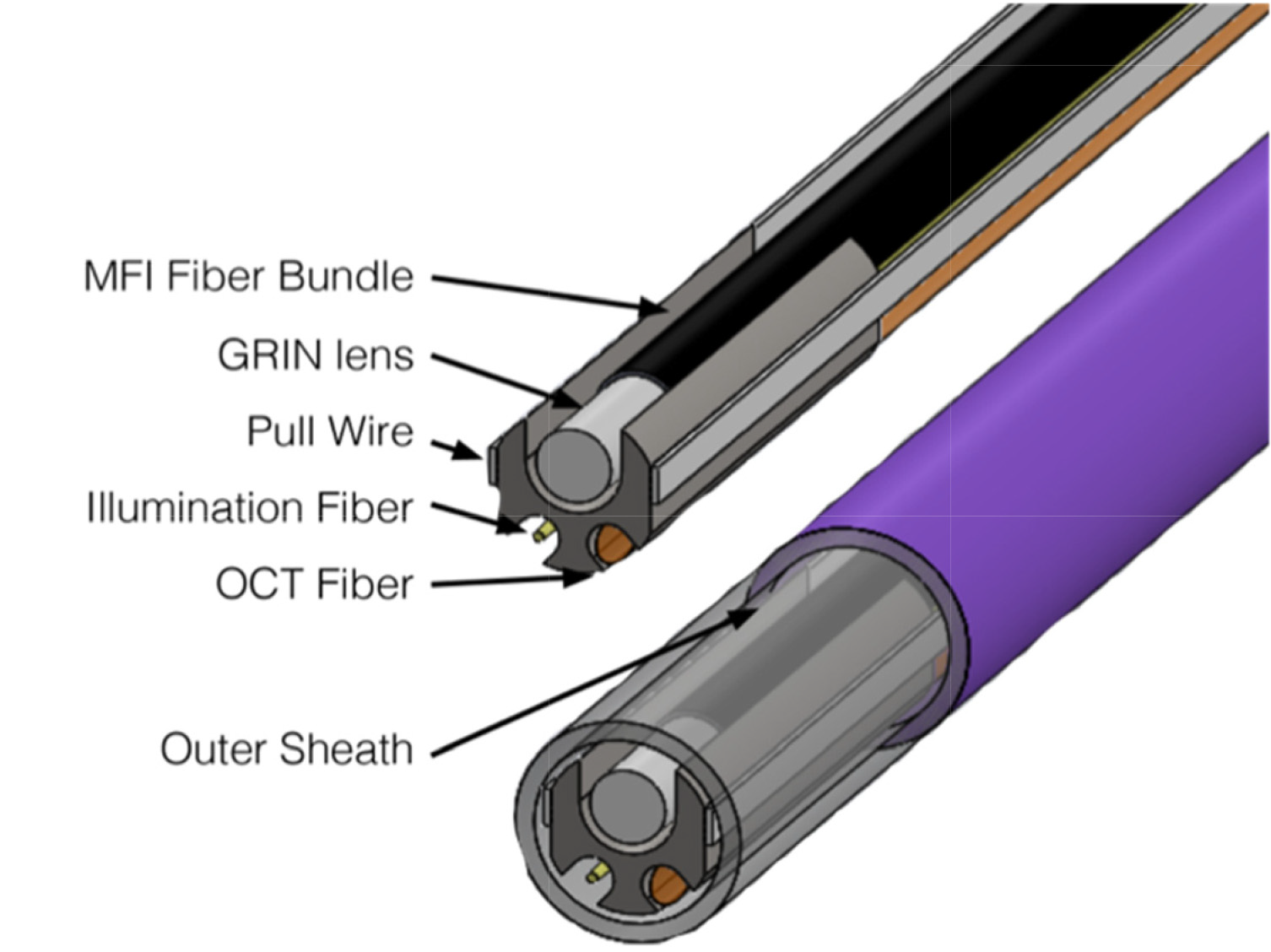
The Falloposcope combines Spectral Domain OCT, pseudo-white light imaging, and fluorescence microscopy into a sub-1mm in diameter endoscope for ovarian cancer detection. Four laser wavelengths are combined into one fiber and sent to the endoscope tip (405nm, 488, 520 nm, and 642 nm). The combination of blue (488nm), green (520nm), and red (642nm) lasers provides pseudo-white light which illuminates the tissue. Light reflected off the tissue is collected by a small lens that is placed at the tip of a 3,000-element fiber. By imaging the other end of the fiber bundle, a real time image of the tissue inside the body can be seen on a computer screen. When the 405nm, 488nm, or 520nm lasers are turned on one at a time and a corresponding filter is placed after the fiber bundle, a fluorescence image is acquired. Each of these wavelengths have been shown to distinguish normal from cancerous tissue in a previous study [1]. Another fiber probe is used for spectral domain OCT. A 1300nm swept source laser is coupled into single mode fiber to propagate the light to the endoscope tip. Small segments of multimode fiber and GRIN (gradient index) fiber are used as miniature versions of spacers and lenses to focus the light onto the tissue. Adding an angle polish to the last piece of fiber creates a mini prism causing the light path to fold making the probe side firing. The resulting OCT signal that makes it back though the fiber probe is analyzed to give a depth image of the tissue. For a 2-dimension image, the physician can manually pull the probe back to get a scan along the tissue. There is a pull wire on either side of the endoscope tip which can be controlled by manipulated by the handle causing one wire to tighten resulting in a bending of the tip of the endoscope. This gives the physician the ability to navigate the endoscope through the fallopian tube to examine any area of interest. A computer mock-up of the endoscope with all the components is shown below [2]:
References [1] T. H. Tate et al., “Multispectral fluorescence imaging of human ovarian and fallopian tube tissue for early-stage cancer detection,” J. Biomed. Opt. 21(5), 56005 (2016) [doi:10.1117/1.JBO.21.5.056005]. [2] Keenan, Molly et al. “Design and characterization of a combined OCT and wide field imaging falloposcope for ovarian cancer detection” Biomedical optics express vol. 8,1 124-136. 8 Dec. 2016, [doi:10.1364/BOE.8.000124] |
 (3)
(3)