About
Kidney Research
|
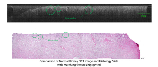
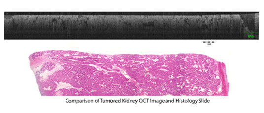
The Tissue Optics Lab has tested the usefulness of OCT in the detection and diagnosis of Renal (Kidney) Cancer. OCT is useful for its very high resolution (2-10 microns), small size, and portability. However the penetration depth of OCT is only about 1-2 mm, limiting the use of OCT imaging to surgical or laproscopic procedures. Because of this, OCT has been streamlined into very small devices that can be implemented in vivo endoscopically. However, for this study, all samples were removed from the body, and viewed ex vivo. Following an IRB-approved protocol, surgical discard tissue was obtained from full and partial nephrectomies of 20 patients. A nephrectomy is the surgical removal of the kidney from the body. For each patient, a sample of normal and diseased kidney was extracted from the removed kidney, each about 1 cm cube in size. These samples were then imaged with an OCT system using a 2 mm endoscope, with a 4 micro axial resolution and 10 micron lateral resolution. The endoscope was placed on top of the sample and a series of images were taken 1 mm apart. Each image was approximately 10 mm lateral and 1.4 mm deep. After imaging, the tissue was fixed in Histochoice, embedded in paraffin and processed by standard protocol to create histology slides. These slides were compared visually to the OCT images to determine matches based on corresponding features that could be seen in both images. Diagnosis was confirmed by a pathologist. Significant difference can be seen between OCT images of normal and cancerous kidney tissue. It is often seen that diseases kidney has disordered artifacts and entirely featureless regions. In normal tissue, normal features such as collecting ducts and glomeruli are visible, unlike in diseased tissue. The ability to differentiate these sections of tissue appears to make OCT useful in verifying clear surgical margins during partial nephrectomies, or other applications where precise identification of cancerous tissue location would be needed. The following pictures give an example of a diseased section of tissue as well as a normal section and display their corresponding OCT image.
|