About
Optical Coherence Microscopy
|
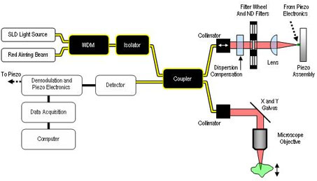
What it is: The optical coherence microscope (OCM) is an optical system that takes depth-resolved en face images of tissue with resolution of approximately 2 µm. OCM uses a technique called interferometry (interference by superposition of two waves). The OCM system detects interference of reference electromagnetic (EM) waves and EM waves scattered from a single plane in the tissue sample. A detector collects the scattered photons to create an image. How it works: The components of the optical coherence microscope system can be categorized into four groups: the light sources, the reference arm, the sample arm, and the data acquisition system. The light sources consist of a super-luminescent diode (SLD) and a red diode laser. The SLD is used for imaging. The SLD has a center wavelength of 841-nm (invisible to the human eye) and a full-width-half-maximum (FWHM) of 48-nm. The red diode laser (visible to the eye) is used to assist the user in positioning the sample. The two light sources are coupled into the same fiber using a Wavelength Division Multiplexer (WDM). The light then travels to a polarization paddle that changes the polarization of the light propagating into the optical isolator. The polarization-dependent isolator insures that no light from the optical system is reflected back to the source. The light then propagates to a two-by-two coupler at which point half of the light is sent to the reference arm and the other half is sent to the sample arm. The reference arm of the OCM is used to modulate the optical path length. The optical path length set in the reference arm of the interferometer defines the location of the en face plane in the sample. The path length can be modulated up to 350 nm using a piezo device. After modulation the light travels back through the reference arm to a two-by-two coupler which sends half the light to the detector and half the light to an optical isolator where it is absorbed. The sample arm directs collimated light onto the en face plane in the sample. X and Y galvanometers are used to direct light and scan the plane of interest. Light is backscattered from the sample and enters a two-by-two coupler. The coupler sends half the light to the detector and half the light to the optical isolator to be absorbed. The light from the reference arm and sample arm are converted to a voltage using a detector and noise if filtered using high pass and low pass filters. The voltages are recorded using a data acquisition system (DAQ board). The computer software maps the voltages at each point in the sample to create a digital image. A schematic of the optical coherence microscope (OCM) is shown in Figure 1-1.
Figure 1-1. Schematic of the optical coherence microscopy system. Advantages of OCM:
The main advantage for using OCM in our lab is that we are able to conduct long term animal studies. Because OCM is non-destructive and can be conducted in vivo, there is no need to sacrifice animals at multiple time points. Long term studies are made easier by using a small diameter rigid endoscope that allows for minimally invasive imaging. Disadvantages of OCM:
|